α-黑素细胞刺激素(aMSH)检测试剂盒(酶联免疫吸附试验法)
ELISA Kit for Alpha-Melanocyte Stimulating Hormone (aMSH)
α-MSH; Intermedins; Alpha-Melanotropin, Alpha-Melanocortin; Alpha-Intermedin
- 编号CEA239Ra
- 物种Rattus norvegicus (Rat,大鼠)相同的名称,不同的物种。
- 实验方法竞争抑制
- 反应时长2h
- 检测范围123.5-10,000pg/mL
- 灵敏度最小可检测剂量小于等于51.9pg/mL.
- 样本类型serum, plasma, tissue homogenates, cell lysates, cell culture supernates and other biological fluids
- 下载英文说明书 中文说明书
- 规格48T96T96T*596T*1096T*100
- 价格¥ 2873¥ 4104¥ 18468¥ 34884¥ 287280
- 欲了解实际交易价格和更多情况,请与当地经销商联系!
 产品包装(模拟)
 产品包装(模拟)
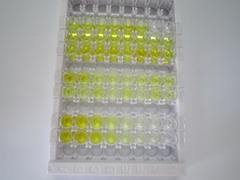 实验结果图
实验结果图
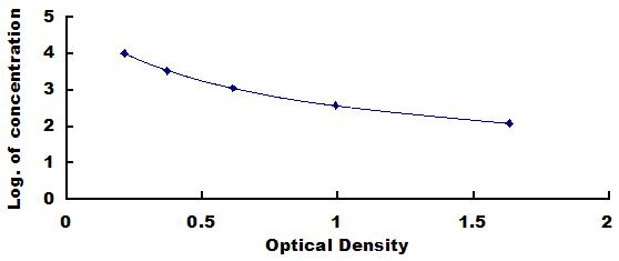 标准曲线图
标准曲线图
 通过ISO 9001、ISO 13485质量体系认证
特异性
本试剂盒用于检测α-黑素细胞刺激素(aMSH),经检测与其它相似物质无明显交叉反应。
由于受到技术及样本来源的限制,不可能完成对所有相关或相似物质交叉反应检测,因此本试剂盒有可能与未经检测的其它物质有交叉反应。
回收率
分别于定值血清及血浆样本中加入一定量的α-黑素细胞刺激素(aMSH)(加标样品),重复测定并计算其均值,回收率为测定值与理论值的比率。
| 样本 | 回收率范围(%) | 平均回收率(%) |
| serum(n=5) | 95-102 | 98 |
| EDTA plasma(n=5) | 88-97 | 93 |
| heparin plasma(n=5) | 96-103 | 99 |
精密度
精密度用样品测定值的变异系数CV表示。CV(%) = SD/mean×100
批内差:取同批次试剂盒对低、中、高值定值样本进行定量检测,每份样本连续测定20 次,分别计算不同浓度样本的平均值及SD值。
批间差:选取3个不同批次的试剂盒分别对低、中、高值定值样本进行定量测定,每个样本使用同一试剂盒重复测定8次,分别计算不同浓度样本的平均值及SD值。
批内差: CV<10%
批间差: CV<12%
线性
在定值血清及血浆样本内加入适量的α-黑素细胞刺激素(aMSH),并倍比稀释成1:2,1:4,1:8,1:16的待测样本,线性范围即为稀释后样本中α-黑素细胞刺激素(aMSH)含量的测定值与理论值的比率。
| 样本 | 1:2 | 1:4 | 1:8 | 1:16 |
| serum(n=5) | 95-104% | 83-93% | 79-88% | 96-105% |
| EDTA plasma(n=5) | 78-99% | 86-93% | 86-94% | 89-96% |
| heparin plasma(n=5) | 89-98% | 78-95% | 89-97% | 87-97% |
稳定性
经测定,试剂盒在有效期内按推荐温度保存,其活性降低率小于5%。
为减小外部因素对试剂盒破坏前后检测值的影响,实验室的环境条件需尽量保持一致,尤其是实验室内温度、湿度及温育条件。其次由同一实验员来进行操作可减少人为误差。
实验流程
1. 实验前标准品、试剂及样本的准备;
2. 加样(标准品及样本)50µL,
加入50µL检测液A(临用前配制);
37°C温育1小时。
3. 洗板3次;
4. 加检测溶液B100µL,37°C孵育30分钟;
5. 洗板5次;
6. 加TMB底物90µL,37°C孵育10-20分钟;
7. 加终止液50µL,立即450nm读数。
实验原理
本试剂盒应用竞争抑制酶联免疫分析法测定标本中待测物质水平。将α-黑素细胞刺激素(aMSH)单克隆抗体包被微孔板,制成固相载体,往包被抗体的微孔中同时加入生物素标记的抗原和待测抗原(标准品或样本),待测抗原与生物素标记抗原对特异性抗体进行竞争结合。温育后经洗涤去掉未结合物,然后加入HRP标记的亲和素,经过温育和彻底洗涤后加入底物TMB显色。TMB在过氧化物酶的催化下转化成蓝色,并在酸的作用下转化成最终的黄色。待测标本浓度越高,标记抗原和抗体的结合就越受到抑制,显色愈浅。显色的深浅与酶量呈正相关,而与样品中待测物质含量呈负相关。用酶标仪在450nm波长下测定吸光度(O.D.值),计算样品浓度。
相关产品
| 编号 | 适用物种:Rattus norvegicus (Rat,大鼠) | 应用(仅供研究使用,不用于临床诊断!) |
| CEA239Ra | α-黑素细胞刺激素(aMSH)检测试剂盒(酶联免疫吸附试验法) | Enzyme-linked immunosorbent assay for Antigen Detection. |
| LMA239Ra | α-黑素细胞刺激素(aMSH)等多因子检测试剂盒(流式荧光发光法) | FLIA Kit for Antigen Detection. |
| KSA239Ra11 | α-黑素细胞刺激素(aMSH)检测试剂盒DIY材料(酶联免疫吸附试验法) | Main materials for "Do It (ELISA Kit) Yourself". |
参考文献
| 杂志 | 参考文献 |
| J Biochem. | Generation of a human bone marrow-derived mesenchymal stem cell line expressing and secreting high levels of bioactive α-melanocyte-stimulating hormone.[Pubmed: 23341471] |
| Pigment Cell Melanoma Res | Pharmacological induction of skin pigmentation unveils the neuroendocrine circuit regulated by light[PubMed: 26582755] |
| The Journal of Biological Chemistry | Intermedin Restores Hyperhomocysteinemia-induced Macrophage Polarization and Improves Insulin Resistance in Mice[Pubmed:27080257] |
| Pigment Cell & Melanoma Research | Pharmacological induction of skin pigmentation unveils the neuroendocrine circuit reCavia (Guinea pig )lated by light[Pubmed:26582755] |
| Nature Communications | Endocrine disruptors induce perturbations in endoplasmic reticulum and mitochondria of human pluripotent stem cell derivatives.[pubmed:28794470] |
| Cell Stem Cell | Super-Obese Patient-Derived iPSC Hypothalamic Neurons Exhibit Obesogenic Signatures and Hormone Responses[Pubmed:29681516] |
| European Neuropsychopharmacology | Epigenetic alterations of the POMC promoter in tobacco dependence[Pubmed:29871818] |
| Neuropeptides | Mechanisms of sustained long-term weight loss after RYGB: α-MSH is a key factor[Pubmed:29685637] |
| Neuropsychobiology | Alcohol Withdrawal and Proopiomelanocortin Neuropeptides in an Animal Model of Alcohol Dependence[Pubmed: 31117084] |
| INTERNATIONAL IMMUNOPHARMACOLOGY | Intermedin alleviates the inflammatory response and stabilizes the endothelial barrier in LPS-induced ARDS through the PI3K/Akt/eNOS signaling pathway[Pubmed: 32892076] |
| J Endocrinol Invest | Serum alpha-melanocyte-stimulating hormone (a-MSH), brain-derived neurotrophic factor (BDNF), and agouti-related protein (AGRP) levels in children with Prader …[Pubmed:35098494] |
